


Murine Anti-mPD-1 mAb
| Anti-mPD-1-mIgG1e3 InvivoFit™ | Unit size | Cat. code | Docs | Price |
|---|---|---|---|---|
RMP1-14-derived mouse monoclonal antibody against murine PD-1 | 1 mg 10 mg (2 x 5 mg) | mpd1-mab15-1 | TDSMSDSDATA | Please contact our distributor Add to favorite |
- About
- Specifications
- Contents
- Details
- Related products
 InvivoGen’s engineeredAnti-mPD-1-mIgG1e3 InvivoFit™ antibody
Recombinant mouse mAb against murine PD-1 for in vivo use
Anti-mPD-1-mIgG1e3 InvivoFit™ is a recombinant monoclonal antibody (mAb) featuring the variable region of the previously described anti-mPD-1 RMP1-14 mAb [1, 2]. The original RMP1-14 hybridoma was obtained by immunizing rats with cells expressing murine programmed cell death 1 (mPD-1; also known as CD279). The use of xenogeneic sequences (i.e. rat origin) for mAbs renders them immunogenic upon injection in mice [3]. Moreover, special attention should apply to mAbs targeting the PD-1/PD-L1 axis, as repeated injections of xenogeneic anti-PD-1 or anti-PD-L1 in tumor-bearing mice were shown to induce fatal hypersensitivity reactions [4]. To overcome this issue, Anti-mPD-1-mIgG1e3 InvivoFit™ was generated by recombinant DNA technology so that it is ~85% murine. It contains the constant region of mouse IgG1 with a D265A point mutation (replacement of aspartic acid by alanine at position 265) resulting in the complete loss of cytolytic effector function [2, 5]. Anti-mPD-1-mIgG1e3 is provided in an InvivoFit™ grade, a high-quality standard specifically adapted to in vivo studies.
Key features of Anti-mPD-1-mIgG1e3 InvivoFit™:
- Sequence is ~85% mouse origin (constant region) and ~15% rat origin (variable region)
- Features the effectorless IgG1e3 (IgG1 with a D265A point mutation)
- Blocks the murine PD-1 receptor without causing T cell depletion
- Guaranteed sterile, endotoxin level < 1 EU/mg
- Specifically designed for in vivo studies in mice
- Low aggregation < 5%
- Produced in both animal-free facilities and defined media
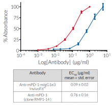
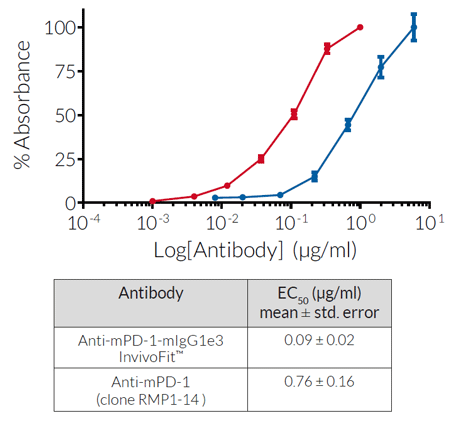
Anti-mPD -1-mIgG1e3 InvivoFit™ is produced in Chinese hamster ovary (CHO) cells, purified by affinity chromatography with protein A, and its binding is validated by flow cytometry and ELISA.
References:
1. Ribas A. & Wolchock J.D., 2018. Cancer immunotherapy using checkpoint blockade. Science. 359:1350-55. 2. Yamazaki T. et al., 2005. Blockade of B7-H1 on macrophages suppresses CD4+ T cell proliferation by augmenting IFN-gamma-induced nitric oxide production. J Immunol. 175(3):1586-92.3. Brüggemann M. et al., 1989. The immunogenicity of chimeric antibodies. J. Exp. Med. 170:2153-2157. 4. Mall C. et al., 2016. Repeated PD-1/PD-L1 monoclonal antibody administration induces fatal xenogeneic hypersensitivity reactions in a murine model of breast cancer. Oncoimmunology. 5(2):e1075114.5. Baudino L. et al., 2008. Crucial role of aspartic acid at position 265 in the CH2 domain for murine IgG2a and IgG2b Fc-associated effector functions. J. Immunol. 181(9):6664-9.
Figures
Specifications
Specificity: Targets cells expressing murine PD-1
Formulation: Lyophilized from 0.2 μm filtered solution in 150 mM sodium chloride, 20 mM sodium phosphate buffer with 5% saccharose
Clonality: Monoclonal antibody
Isotype: Murine IgG1e3 (D265A mutation; no effector function), kappa
Source: CHO cells
Purity: Purified by affinity chromatography with protein A
Tested applications: Flow cytometry and ELISA
Quality control:
- Binding confirmed by flow cytometry
- The complete sequence of this antibody has been verified
- < 5% aggregates (confirmed by size exclusion chromatography)
- Guaranteed sterile and <1 EU/mg (determined by the LAL assay)
Contents
Anti-mPD-1-mIgG1e3 InvivoFit™ is provided sterile, endotoxin-free, azide-free and lyophilized.
This product is available in two pack sizes:
- mpd1-mab15-1: 1 mg
- mpd1-mab15-10: 10 mg (2 x 5 mg)
![]() Product is shipped at room temperature.
Product is shipped at room temperature.
![]() Store lyophilized antibody at -20 °C.
Store lyophilized antibody at -20 °C.
![]() Lyophilized product is stable for at least 1 year
Lyophilized product is stable for at least 1 year
![]() Avoid repeated freeze-thaw cycles.
Avoid repeated freeze-thaw cycles.
Details
Programmed cell death 1 (PD-1; also known as CD279) is a type I transmembrane protein expressed at the cell surface of activated and exhausted conventional T cells. PD-1 is an inhibitory immune checkpoint that prevents T-cell overstimulation and host damage.PD-1 interaction with its ligands PD-L1 (programmed cell death ligand 1) or PD-L2 induces inhibition of T-cell receptor signaling. Blockade of PD-1 with mAbs has allowed unprecedented remissions in patients with metastatic melanoma or non-small cell lung cancer.
Back to the topYou may also need
Raji-hPD-1 CellsHuman lymphoblast cells - ADCC PD-1 Target Cells
Anti-mCTLA-4Antibody against murine CTLA-4
Anti-hPD1 isotype familyFamily of antibodies against human PD-1
Mouse IgG1e3 Controlβ-galactosidase antibody (isotype control) - Mouse IgG1e3